
This post is also available in: polski (Polish)
The design of the prototype of the e-Medicus application based on the level set functions and computational intelligence algorithms.
INTRODUCTION
The planned solution will be based on the latest scientific achievements in the field of image segmentation, computational intelligence algorithms, mathematical models and theories. The system will be responsible for registering and analyzing medical images in the form of numerical functions. This will allow for the identification of medical changes and appropriate data classification. The multimedia presentation of the process of changes will be carried out with the use of the level set function, which enables the topological change of the properties of the solution. The project will develop and apply new procedures and algorithms in the field of theoretical computer science and numerical mathematics using the issues of neural networks, genetic algorithms, semantic networks, image ontology, rough set theory, level set methods and hybrid algorithms. The designed algorithms will develop methods and concepts in the field of image segmentation, capturing, transferring, collecting and extracting information and they will present them in the appropriate form.
THE MODEL OF THE SYSTEM
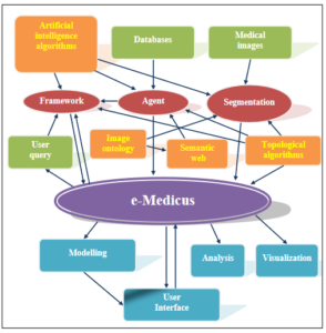
The e-Medicus system consists of:
- artificial intelligence algorithms
- agents (bots)
- segmentation algorithms
- framework,
- warehouses / databases
- visualization system,
- adaptive user interface.
 DIGITAL RADIOGRAPHY
 DIGITAL RADIOGRAPHY
The medical images, which for more than one hundred years have been recorded in analogue form on X-ray film, have recently become the subject of the intense research. The X-ray film allowed for the recording and presentation of the image as well as its storage. At present radiovisiography, which was initially used only in dentistry, has become the standard. With time, however, despite of the fact it created much controversy, it appeared in other fields of medicine.
The images created in this standard are saved in digital form as a two-dimensional matrix, each element of which constitutes the value that determines the grayscale level of the respective pixel. Digital recording is a great advantage of this method, because it allowed for easier storage of images and their possible transmission for further diagnosis. Moreover, standard software provided with radiovisiography equipment offer basic image operations, such as changing the image presentation manner (rotate by any angle, zoom in or zoom out), changing the contrast or brightness, making any type of measurements (eg. angle and surface area measurements). In addition, the high quality of the sensor significantly reduces the radiation doses to which the patient is exposed during the examination. However, it remains unchanged that diagnosis of tissue anomalies is still the responsibility of the physician. Due to this, along with the development of technology for creating medical images, software supporting their analysis is created. .
ANALYSIS OF MEDICAL IMAGES
The process of computer image analysis starts at the level of pixels. The image is presented in a form of two-dimensional pixel array providing only information about the location and color of individual points of the image, but does not contain information determining which pixels create particular objects. In order to analyze an image, it is necessary to go from pixel level to object level. It is done with help of less or more complex segmentation algorithms.
MEDICAL IMAGES SEGMENTATION
Among the simplest segmentation methods, one should mention segmentation by thresholding and by detecting object edges. The first one consists in determining a certain threshold value T, on the basis of which each image pixel is assigned to one of the two categories. Multilevel thresholding is also possible. In turn, the second mentioned method is based on the boundaries between the areas of different brightness, where a large difference between the levels on the grayscale of the adjacent pixels indicates the presence of edges between the objects.
SEGMENTATION USING STATISTICAL METHODS
Presented above algorithms were used to look for soft tissues lesions in the dental region. Figures 1 and 2 show the results of their application. It is visible that with a properly selected number of classes and the method of measuring the distance between the points, they are very similar.

Figure 1 Image segmentation into five clusters with k-means algorithm using the Euclidean distance.

Figure 2 Image segmentation with the 4-nearest neighbors algorithm using the Euclidean distance
SEGMENTATION BASED ON THE LEVEL SET METHOD
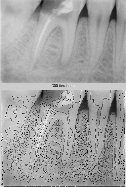
Figure 3 Image segmentation with the algorithm of the variational level set method after 300 iterations and the level set function initiated at a distance of 6 pixels from the edge of the area.
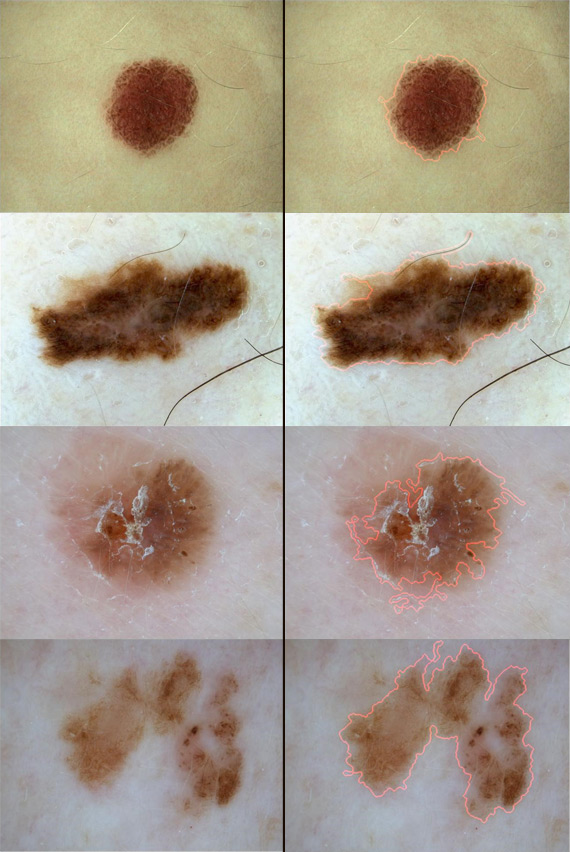
Figure 4 Dermatoscopic image segmentation with use of the level set method
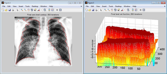
Figure 5 Image reconstruction using the variational level set method
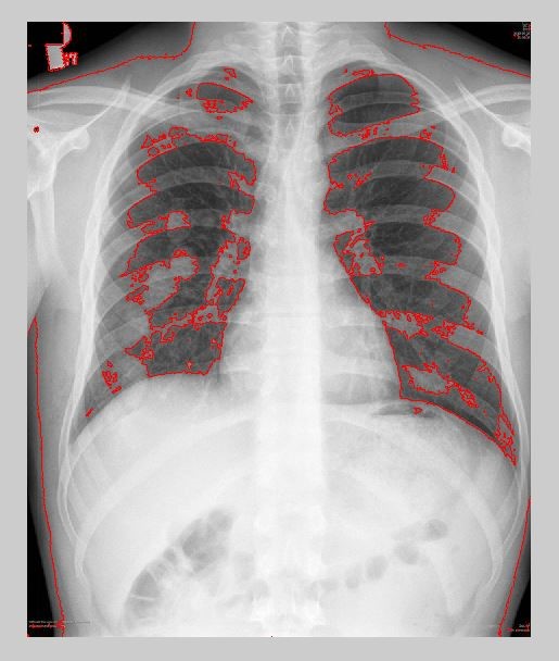
Figure 6 Image reconstruction using the modified level set method
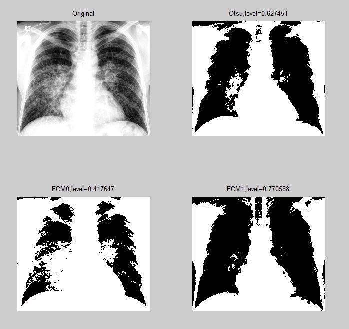
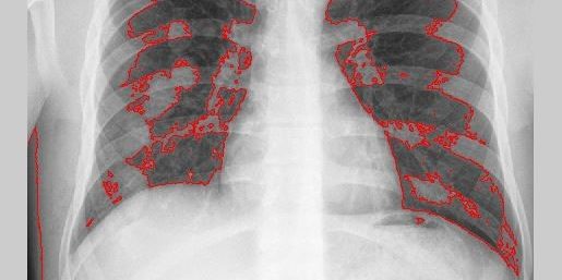
Figure 7 FCMThresholding – sarcoidosis
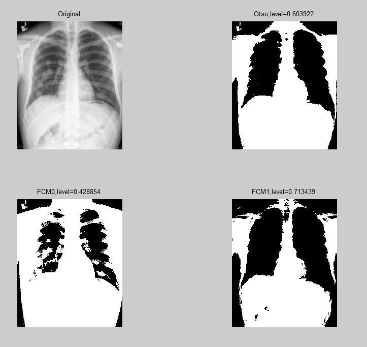
Figure 8 FCMThresholding – metastases
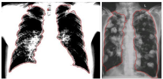
Figure 9 Results of the contour function. Sarcoidosis in the image on the left, metastasis on the right.
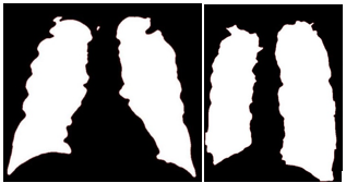
Figure 10 Results of the contourf function. Sarcoidosis in the image on the left, metastasis on the right. The function divides the image into two areas – black and white, where the white area is the area that we are interested in for further analysis.
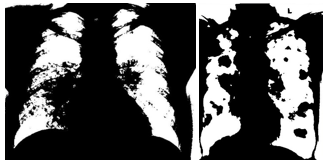
Figure 11 Sarcoidosis – image on the left, metastasis – image on the right.
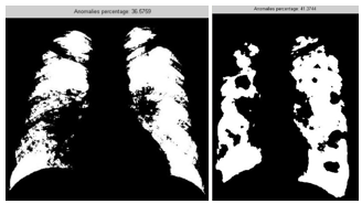
Figure 12 Final results. Estimated value of the anomaly. The results are shown in each picture, the left one shows the results of sarcoidosis and the right one shows the metastasis.